
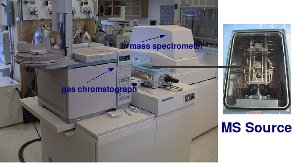
Computerized gas chromatography – mass spectrometry (GCMS) is the principal method used to detect and identify compounds by exploiting the mass variations of different compounds. A typical GCMS system performs six functions:
- Compound separation by gas chromatography
- Transfer of separated compounds to the ionizing chamber of the mass spectrometer
- Ionization
- Mass analysis
- Detection of the ions by the electron multiplier
- Acquisition, processing and display of data by computer
-
Compound separation by gas chromatography
A syringe injects a known amount of the saturate fraction dissolved in hexane into the GC column, which is a long, thin capillary tube 0.32 mm wide and 60 metres long. The inner surface of the column is coated with a film of nonvolatile liquid, called the stationary phase. The temperature of the column is gradually raised, using a temperature-programmed oven, causing the compounds to move. Each injected sample is vaporized and mixed with helium, an inert carrier gas, which is also called the mobile phase.
The larger molecules are retained by the stationary phase at the head of the column, a process called ‘cold trapping’. The mobile phase and sample mixture move along the column, while components are separated as they are repeatedly retained by the stationary phase and released into the mobile phase depending on their volatility and affinity for each phase. The compounds elute, or are washed out from the end of the column, at different rates and are analyzed by the mass spectrometer.
-
Transfer of separated compounds to the ionizing chamber of the mass spectrometer
-
Ionization
This step is the beginning of mass spectrometry. There are several modes of ionization, including electron impact (EI) ionization, chemical ionization (CI) and field ionization. Generally, CI provides information on the molecular weight of the compound. EI yields specific information on fragments ions that help to specifically identify the organic compound. Here, we will go into detail about EI.In EI ionization, the eluting compounds pass directly from the GC column to the ionizing chamber (or source, see picture at the top of this page) of an MS where they are ionized by an electron beam. Each molecule (M) eluting from the GC is bombarded with energetic electrons, which cause it to form molecular ions (M.+) as follows:
M + e– → M.+ + 2e–
The molecular ion can undergo further fragmentation or rearrangement to form other ions, neutral molecules or radical ions. Fragment ions are electrically charged dissociation products from a parent ion. They may dissociate further to form other electrically charged molecular or atomic fragments (also called moieties) of successively lower formula weight. The fragment ions generated in the ion source chamber are accelerated toward the detector through the mass analyzer by a differential voltage.
-
Mass analysis
In the mass analyzer, the ions are concentrated into a beam so that only positive ions of a given mass/charge (m/z) ratio hit the detector at any moment (thus, the neutral molecules are not detected). The two main methods to concentrate and analyze the ion beam use either magnets or quadrupole rods. Our machines use magnets.
-
Detection of the ions by the electron multiplier
Using an electron multipler during a typical scan analysis, the detector measures ions over the mass range m/z 50 to m/z 600 every three seconds. Because many lives and much, much science is produced through the analysis of mass spectral data, we decided to dedicate a page to the principles of the scan analysis, the output of a GCMS.

-
Acquisition, processing and display of data by computer
During a typical scan run, which lasts ~90 minutes, where the mass spectrometer scans the mass range of interest every three seconds, 1800 mass spectra are generated for each sample ([90 min * 60 sec/min]/ 3 sec/scan). Such large amounts of data require data systems for storage and manipulation.
There are several operating modes for GCMS, which help to give us different and more specific information about our organic compounds.
In addition to identifying and quantifying compounds present in a sample, we may also want to know its isotopic composition, which we find through compound-specific isotope analysis.