
Lipids from modern microbial communities or cultures often contain highly polar moieties or have other properties that render them nonvolatile, and not amenable to analysis by GC-MS without derivatization, degradative wet chemistry, or pyrolysis.
LC-MS (liquid chromatography – mass spectrometry) is an alternative analytical technique that allows us to examine intact polar lipids (IPLs) without employing these degradative methods. To see a couple of comparative reactions of degradative methods vs. LC-MS, go here.
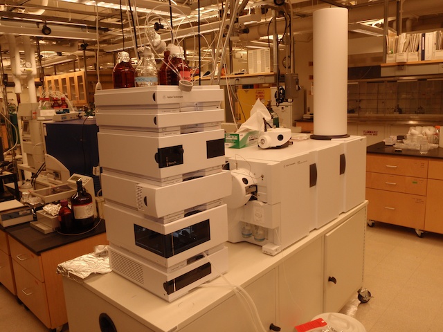
The system we use is composed of two units: The first, High Pressure Liquid Chromatography (HPLC) separates the compounds by passing them through a silica-based column that contains a bonded stationary phase selected to optimize separation of various compound classes. Compounds are eluted by a liquid mobile phase – usually an organic solvent, water, or a mixture. The HPLC can also be used preparatively, in some cases we can separate pure compounds and subject them to further analysis such as NMR or radiocarbon analysis.
The second unit is the detector, typically a mass spectrometer. Before compounds can be detected via mass spectrometry they need to be ionized first. The ionization source therefore comprises the interface between the HPLC and the mass spectrometer. We use both electrospray ionization (ESI) and atmospheric pressure chemical ionization (APCI) for polar lipid analysis. Both types of ionization occur under atmospheric pressure in a nitrogen atmosphere. ESI is a soft ionization technique used for the ionization for more polar compounds such as IPLs. It operates by applying a strong electric field to a capillary containing the liquid mobile phase and analytes from the HPLC. The field causes charge to accumulate on the surface of the liquid, which breaks into small droplets (the ‘spray’). As the droplets break apart the charge is desorbed from the droplet to the analyte. APCI is also a soft ionization technique that is used for less polar compounds, such as glycerol dialkyl glycerol tetraethers (GDGTs) and bacteriohopanepolyols (BHPs). As solvent moves into the APCI source it is heated to a high temperature, such that both the mobile phase and analytes are gaseous by the time they encounter the corona discharge electrode. The corona discharge forms primary ions of N2+, which reacts with the evaporated mobile phase to form secondary ions which then react with the analyte. ESI and APCI can be selected for positive ion or negative ion modes, which give complementary structural information.
In our lab we use two types of mass spectrometers to detect and identify lipids, an Agilent Technologies 6130 SingleQuad and an Agilent Technologies Accurate-Mass Q-TOF 6520. Our SingleQuad system is equipped with a mass selective detector (MSD) that can be operated in selective ion monitoring (SIM) mode which allows for very sensitive detection of analytes and a multiple wavelength detector (MWD) to analyze pigments. Additionally our SingeQuad systems can be operated in preparative mode using time based or peak based fraction collection. The QTOF is a mass spectrometer with two mass analyzers consisting of a quadrupole ‘front end’ (Q) and a time of flight (TOF) and the second mass analyzer. The analyzers are separated by a collision chamber. This means that we can do true MS-MS experiments, using the quadrupole to select a precursor mass of interest (for example the molecular ion of an IPL) then applying a voltage to the collision cell causing fragmentation, then using the TOF to detect the product ions produced. Additionally, the TOF has a mass precision of <2 mDa, allowing us to determine unique elemental composition by the exact mass of a compound.
References
- Talbot H.M., Summons R.E., Jahnke L.L. and Farrimond P., 2003. Characteristic fragmentation of bacteriohopanepolyols during atmospheric pressure chemical ionisation liquid chromatography/ion trap mass spectrometry. Rapid Communications in Mass Spectrometry 17, 1-9.
- Sturt H.F., Summons R.E., Smith K., Elvert M. and Hinrichs K.-U., 2004. Intact polar membrane lipids in prokaryotes and sediments deciphered by ESI-HPLC-MS n New biomarkers for biogeochemistry and microbial ecology. Rapid Communications in Mass Spectrometry 18, 617-628.
- Liu X.-L., Summons R.E. and Hinrichs K.-U., 2012. Extending the known range of glycerol ether lipids in the environment: structural assignments based on MS/MS fragmentation patterns. Rapid Communications in Mass Spectrometry 26, 2295–2302.
- Schubotz F., Meyer-Dombard D.R., Bradley A.S., Fredricks H.F., Hinrichs K.-U., Shock E.L., and Summons R.E., 2013. Spatial and temporal variability of biomarkers and microbial diversity reveal metabolic and community flexibility in Streamer Biofilm Communities in the Lower Geyser Basin, Yellowstone National Park. Geobiology 11:549-569.